Originally published in 1895, the article below describes what it was like inside a dissecting room. What I found interesting was the description of how the bodies were prepared for dissection. This information can be found in the latter half of the article.
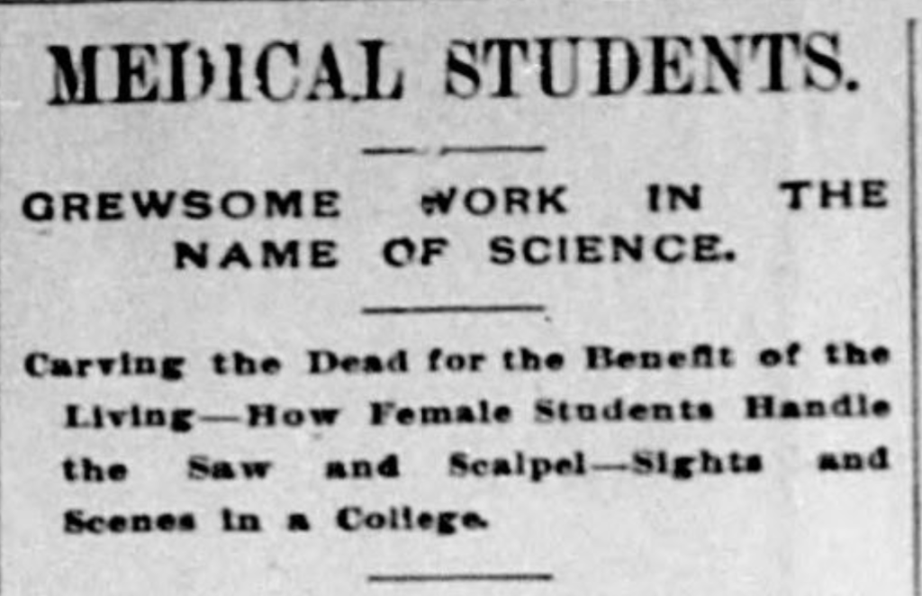
Medical Students
Gruesome Work in the Name of Science

Man’s usefulness does not always end with death, nor does every corpse return to the traditional dust. Even the most worthless of men in life become valuable after death – to medical students at least.
There are several places in Omaha where dead bodies are put to scientific use. These places are called dissecting rooms, and are generally connected with some of the medical colleges, but occasionally a student gets a subject that is particularly interesting and takes it to his own office for dissection.
A Bee reporter managed to secure admission to the dissecting room of one of Omaha’s medical schools one night recently, where he was permitted to witness the work in full operation, of cutting up corpses. The room was in the top story of a large building and to all outside appearances there was nothing going on inside. Upon opening the door a strong flood of light poured forth, as well as a stench which was strong enough to take its own part. In fact, it caused the reporter to become afflicted with a tremendous desire to imitate the whale which swallowed Jonah. The door was closed and the visitor found himself in the company of a baker’s dozen of corpses in various stages of decomposition and dissection. A number of students were hard at work cutting and slashing the bodies in the interest of science, while the reporter was hard at work trying to put a stop to the internal strife which seemed to be gaining strength in his digestive organs. The students were too busy to pay much attention to him, however, and a strong cigar helped him to retain his last meal.
All over the room were long, narrow tables on which were the “stiffs.” The students worked over and leaned on these bodies with the utmost familiarity, and discussed the different formations of each. Over in one corner a student was studying the brain of a subject, while another was learning the muscular parts of an arm. One students was slashing into the abdomen of a large-sized man, while one was discussing a spinal nerve system with a classmate.
A pale-faced young woman with intellectual brow and bloody hands was examining the muscular action and formation of the heart and lungs of a small-sized corpse, while one of the professors was discussing, in a learned manner, the best method for performing a difficult case of surgery. The young woman looked as if she might faint at the sight of a mouse or a bloody nose, but she went at her work with a decided relish, and she cut and slashed with a keen knife as if she enjoyed it. Another young woman was assisting her, and it was afterward learned that these two females were the most advanced students in their class. They were great students, and were able to practically demonstrate the lessons obtained from medical journals.

Soon other students appeared, and in a short time some one was working on each of the dozen corpses in the room. A stout, strong-looking man opened a trap door, lowered a block and tackle, and in a few minutes another corpse was hauled up, apparently from under the floor. This one was put upon a table and prepared for operation by having the location of the internal organs outlined upon the skin. A couple of first-year students were given a chance to carve these remains. The room presented a busy appearance, young men dressed in rubber coats or old clothes, and armed with sharp knives were cutting away flesh and skin, carefully exposing the muscles and nerves, performing difficult operations by proxy, as it were, and engaging in comparing their work with the subjects which they were studying in books. An exclamation from one quiet young man brought others to his side. He exposed the vermiform appendix of the “stiff” over which he was working, and in it was a grape seed. The first symptoms of inflammation were noticeable, and he was of the opinion that in a few days a well developed case of appendicitis would have been the result. He bemoaned the fact that the man had died suddenly without giving the appendix a chance to get in its deadly work, and so did his fellows. The appendix looked like a long white string, but none of the students could give any reason for its existence in the human body.
He explained that when a “stiff” was brought to college for dissection a half pound of arsenic was injected into the body, thoroughly disinfecting it and preserving the tissues. After a few days liquid starch, colored with aniline, was forced into the arteries and veins making them assume a natural appearance. After the muscles, nerves, internal organs, skin, and ligaments had been removed from the subject the bones were boiled in vats of acid, removing every particle of matter clinging to them. Then they were bleached and string together on wires, giving each of the graduates a skeleton to hang in his own closet. The more remarkable subjects were duplicated in wax and preserved for the lecture room.
There is no doubt but that surgery and medical practice has made rapid strides within the last decade and it is no uncommon thing to see old practicing physicians and surgeons become students in the advanced classes in this medical college in order to keep up with the advancements made in their profession.
Source: The Lamar register. (Lamar, Colo.), 02 Feb. 1895.

